Facilitation of wound closure through edge coping with the help of Deep Oscillation®: A Case Report by Diana M. Miranda Monge. TF

Diana M. Miranda Monge. TF Facebook Instagram
Abstract
Treatment for the closure of the wound due to deposition after skull surgery for subdural haematoma, placement, removal and re-placement of a cranial prosthesis. Due to its aetiology, the wound cannot heal from the bottom up due to the presence of the prosthesis, therefore, it can only heal from the edges towards the centre. Application of Deep Oscillation® for the adequate management of the wound edges, decrease of fibrosis and increase of the tensegrity of the skin to be able to cope and achieve closure.
Keywords: Wound dehiscence, skull, fibrosis, deep oscillation, edge coping, cranial prosthesis.
Introduction
Wound healing is a complex process in which a significant number of cells interact in a specific way, carrying out a sequence to repair the injured tissue (1). It is a fascinating mechanism that provides an evolutionary advantage until the wound is closed and thus ensures the survival of the individual, creating a new capillary bed through angiogenesis, being initially created under a not-so-structured form of tissue and later arriving at normal skin density and composition, resulting in scar tissue (1,2).
The process as such basically consists of consecutive stages: haemostasis (clot formation) (5), inflammation (3,5), proliferation (formation of granulation tissue) (3,5), and tissue remodelling (maturation) (5), however, there are two mechanisms that seem to be involved, which are intrinsically related, which are inflammation and proliferation (3), which leads to the deduction that in a chronic lesion with a mild inflammation response, it is less likely to conclude that there are relevant factors that help to complete the tissue repair process, causing a stagnation of the injury itself.
There are implications in the management of injuries that make it extend towards a multidisciplinary approach, in which the Physical Therapist is positioned as an important element in the proper management of wounds, producing optimal results in treatment (4), through the early detection of tissue injuries, wound management from a rehabilitative point of view, and prevention of integumentary injuries (4).
As part of this case study, the physiotherapeutic intervention of the use of Deep Oscillation® in a wound dehiscence (9) located at the level of the skull (8) after the placement of a prosthesis will be presented. Given the chronicity of the lesion, factors such as local blood supply, immune response, underlying diseases that may delay healing, medications, extrinsic factors such as pressure on the lesion area, humidity and temperature management must be taken into account (6) .As part of the specific approach, the wound must be classified within the assessment scales according to its stage (7), prepare the bed, and in this case what was worked on most was the state of the edges, since granulation could not be reached from the bottom up due to the presence of the prosthesis, if not that only a proliferation of the tissue of the edges could be achieved towards the centre, which were with fibrosis, which made it impossible to cope with them due to the alteration of the architecture and normal function of tissues (10).
Case Report
A 68-year-old man is admitted to the hospital on 1/2/2019 with a traumatic subdural haematoma (after a hit from a soccer ball) in the left lobe of the brain, he underwent a craniotomy for lavage plus drainage of the haematoma. On 2/12/19, an extension of the craniotomy plus drainage and lavage of the haematoma (residual fluid) was performed. Again, on 2/18/19, the craniotomy was reopened to wash out the encapsulated infection (not meningitis), this time the part of the skull could not be placed again due to the presence of significant inflammation at the brain level.
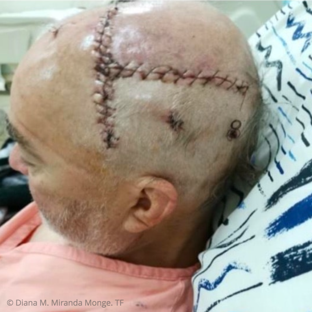
Figure 1: Cranial surgery
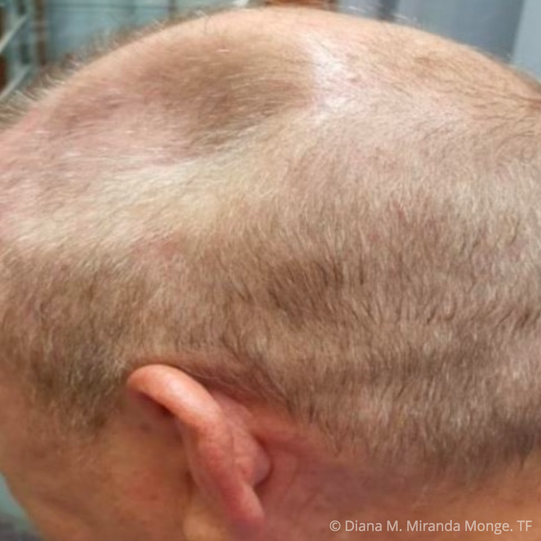
Figure 2: Removal of the cranial prosthesis..
As a result of the cranial collapse, on 7/28/19 he had a seizure and was admitted again. On 8/5/19 a cranioplasty was performed. Subsequently, on 4/28/21, he presented a significant exudate and as a result, a dehiscence of the wound appeared in part of the scar that had been gradually sinking.
The use of hydrogel was recommended as a treatment by hospital staff for wound management.
As of 06/10/2021, physiotherapeutic treatment begins, the wound bed is assessed and it is determined that there is good humidity control, there is no presence of granulation tissue due to the placement of the cranial prosthesis, therefore, its closure must be carried out by approximation of the edges, and even though they are undercut and fibrosed, the tension of the skin is strong and there is very little mobility of the edges of the wound.
It begins with the placement of a transparent sterile dressing over the lesion to perform the application of the Deep Oscillation, during the first two sessions the following sequence was used:
Haematoma: 170Hz - 200Hz 10 minutes, 15Hz - 28Hz 5 minutes, 70Hz - 100Hz 5 minutes
Fibrosis: 160Hz - 180Hz 15 minutes, 60Hz - 100Hz 5 minutes
Placement of hydrogel, collagen and proceed to keep the wound covered with sterile gauze.
Subsequently, the sequence was worked on:
Fibrosis: 160Hz - 180Hz 15 minutes, 60Hz - 100Hz 5 minutes
The applications were carried out every three days until 07/24/2021, where it was possible due to the change in the tensegrity of the wound edges due to the effects of Deep Oscillation, to carry out a mechanical coping with surgical tape, to then put on a collagen dressing.
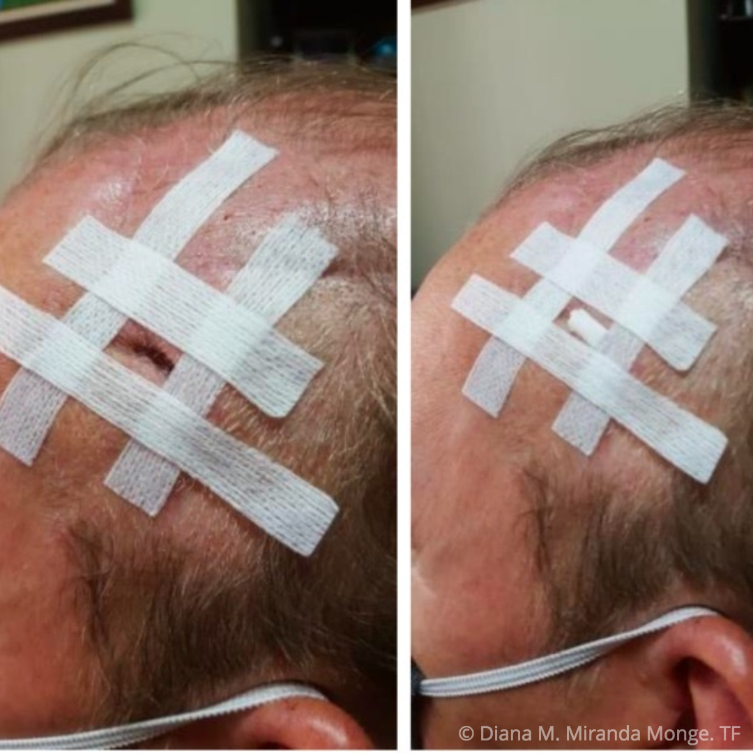
Figure 3: Edge coping with surgical tape
In the next session on 7/28/2021 these were the results obtained:
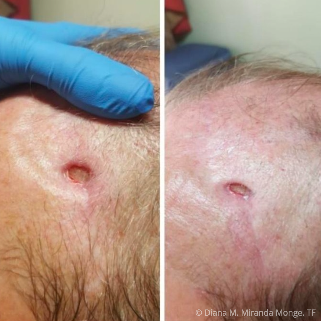
Figure 4: Final result
Today we continue with the coping of edges and the use of a biocellulose dressing to conclude with the closure of the wound.
Discussion
Fibrosis at the edges of wounds can cause a delay in the wound closure process due to the fact that the development of cells in the bed becomes irregular and chronic (10), this type of fibrotic tissue alters the normal function of the tissues, thus, it is necessary to bring the lesion to a point where a controlled process of regenerative activities is stimulated, with an increase in the substances that generate adequate angiogenesis.
Given this process, the use of Deep Oscillation® is used, supported by studies that indicate its use as an adjunctive treatment option so that the resonance vibrations favour the transport of interstitial fluid and other substances to the lesion bed, favouring the mechanical activation and drainage in the area to be treated with the benefit of dissolving fibrosis present in the tissue (11) and improving the tensegrity of the perilesional skin.
While the wound is still in its repair process until its closure, it requires nutrients and oxygen, so the control of oedema and humidity within it plays a fundamental role (9), metabolic un-controls can cause a delay in healing as well as the aforementioned factors, without neglecting the potential of the wound to go from a state of colonization to one of infection, therefore, it is crucial to take into account any change in the state of this both locally and at the systemic level, since these changes imply prevention in the appearance of adverse circumstances that could interrupt the process that is gradually being carried out.
Conclusions
In summary, dehiscence in wounds is an aspect that can be prevented through the necessary care in the postoperative period, reducing risk factors that can cause this situation, however, when faced with a scenario like this, all should be assessed the aspects that lead the wound bed to be in optimal conditions to be able to have a successful healing and to be able to have the perilesional area in the best possible state, since it is essential to achieve the objective of closure with resistant, strong tissue and with good mobility.
Bibliographic references
1. Clough BA, March S, Chan RJ, Casey LM, Phillips R, Ireland MJ. Psychosocial interventions for managing occupational stress and burnout among medical doctors: a systematic review. Syst Rev [Internet]. 2017;6(1).
2. Sorg H, Tilkorn DJ, Hager S, Hauser J, Mirastschijski U. Skin Wound Healing: An Update on the Current Knowledge and Concepts. Eur Surg Res. 2017;58(1- 2):81-94. doi: 10.1159/000454919. Epub 2016 Dec 15. PMID: 27974711
3. Kasuya A, Tokura Y. Attempts to accelerate wound healing. J Dermatol Sci. 2014;76(3):169–72.
4. Moore KD, Hardin A, VanHoose L, Huang H-H. Current wound care education in entry-level doctor of physical therapy curricula. Adv Skin Wound Care. 2020;33(1):47–52.
5. Rezaie F, Momeni-Moghaddam M, Naderi-Meshkin H. Regeneration and repair of skin wounds: Various strategies for treatment. Int J Low Extrem Wounds. 2019;18(3):247–61.
6. Han G, Ceilley R. Chronic wound healing: A review of current management and treatments. Adv Ther. 2017;34(3):599– 610.
7. Goldberg SR, Diegelmann RF. What makes wounds chronic. Surg Clin North Am. 2020;100(4):681–93.
8. Chen Y-W, Shih C-T, Cheng C-Y, Lin Y-C. The development of skull prosthesis through active contour model. J Med Syst. 2017;41(10):164
9. Doughty DB. Preventing and managing surgical wound dehiscence. Home Healthc Nurse. 2004;22(6):364–7.
10. Maria ATJ, Bourgier C, Martinaud C, Borie R, Rozier P, Riviere S, et al. De la fibrogenese a la fibrose : mechanismes physiopathologiques et presentations cliniques. Rev Med 2020;41(5):325–9.
11. Interne. Boisnic S, Branchet M-C. inflammatory and draining effect of the Deep Oscillation® device tested clinically and on a model of human skin maintained in survival condition. Eur J Dermatol. 2013;23(1):59–63