
A PhysioPod® UK Independent Reflection: Tissue-Centred Lipoedema Care
Lipoedema is too often reduced to a simple "weight problem" or judged purely by its outward, visual staging. This reflection takes a different path. It is a comprehensive translation of the latest clinical science—exploring everything from the genetics of adipose heterogeneity and the metabolic effects of GLP-1s, to the physical mechanics of joint displacement and the deep-acting biophysics of Deep Oscillation.

- Introduction to tissue-centred clinical reasoning
- Lipoedema as a chronic, systemic loose connective tissue pathology
- Where is lipoedema fat found?
- The non-progressive nature of lipoedema
- The evolution of lipoedema science, advocacy, and clinical milestones (1940–2026)
- Nutrition and weight management: protecting the lymphatic system
- Tissue-centred care: treating what the tissue needs today
- How Deep Oscillation interacts in the tissue layers
- Supporting connective tissue: direct and extrapolative clinical evidence
- Surgical integration: supporting pre- and post-operative pathways
- Combining therapies for daily life
- Clinical glossary of biophysical and medical terms
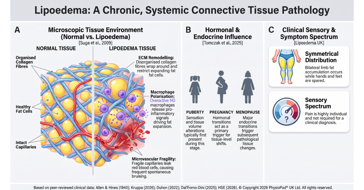
- Figure 1: Lipoedema connective tissue pathology
- Figure 2: Anatomical distribution & sparing
- Figure 3: Why BMI Fails in lipoedema: reframing progress through WHtR
- Figure 4: The hypersensitive tissue state
- Figure 5: The congested tissue state
- Figure 6: The indurated tissue state
- Figure 7: Mechanobiology of Deep Oscillation in human tissue layers
- Figure 8: Deep Oscillation in the surgical pathway
- Video 1: Deep Oscillation theory and visual evidence
1. Introduction to tissue-centred clinical reasoning

Dr Solangel Hernández Tápanes, PhD, MS
2. Lipoedema as a chronic, systemic loose connective tissue pathology
The Somatic and Mechanical Reality: Gravity, Joint Displacement, and Pain Modulation
3. Where is lipoedema fat found?
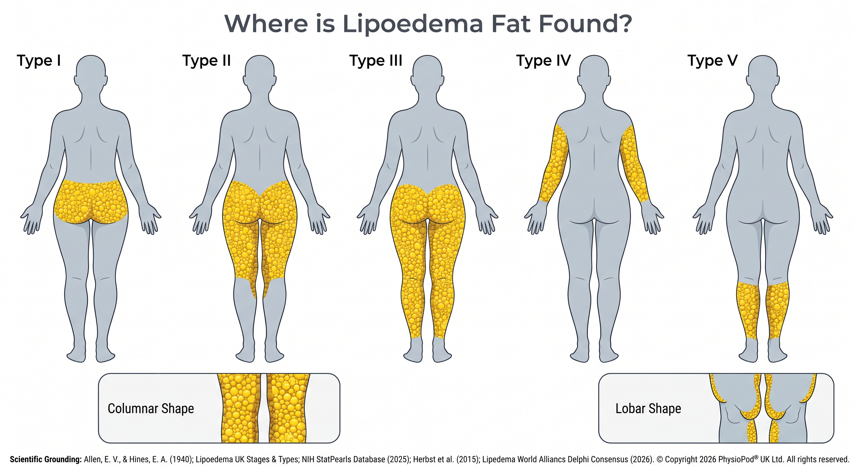
Figure 2: Anatomical distribution & sparing - Clinical mapping of the five anatomical types of lipoedema adipose distribution. © Copyright 2026 PhysioPod® UK Ltd. All rights reserved.
A. The five anatomical types and shape descriptors
-
- Type I: Pelvis, buttocks, and hips (often referred to as the "saddlebag" phenomenon).
- Type II: Buttocks to knees, frequently accompanied by prominent folds of fat forming around the inner side of the knees.
- Type III: Buttocks to ankles.
- Type IV: Arms.
- Type V: Lower legs (isolated to the calf and ankle area).
-
- Columnar shape: Symmetrical enlargement of the lower limbs which become column-shaped, cylindrical, or trunk-like, losing natural contour.
- Lobar shape: The presence of large, prominent bulges or lobes of fat overlying the lower extremities, hips, or upper arms.
B. The "sparing" rule and diagnostic cuffing
- In lymphoedema, swelling typically includes the feet and hands, presenting with a positive Stemmer sign (the inability to pinch a fold of skin at the base of the second toe).
- In lipoedema, because the hands and feet are typically spared, patients present with a negative Stemmer sign.
C. Compassionate clinical reasoning and widespread tissue distress
When patients living with lipoedema share their experiences on forums like the active Reddit r/lipedema community, they often describe a distressing reality: feeling the condition "everywhere." For the individual, this represents a profound and deeply valid physical experience. Within these digital networks, women frequently report discovering painful, lumpy tissue or firm nodules in unexpected areas, such as a stomach that suddenly feels "lumpy," or experiencing a sudden sensation of the condition "exploding everywhere" following major hormonal shifts. Rather than representing an exaggeration, these shared physical accounts are highly consistent with the systemic nature of this loose connective tissue pathology.
4. The non-progressive nature of lipoedema
5. The evolution of lipoedema science, advocacy, and clinical milestones (1940–2026)
6. Nutrition and weight management: protecting the lymphatic system
Key clinical findings demonstrate:
- The waist-to-height ratio (WHtR) replaces the scales: Since lipoedema fat is highly resistant to traditional calorie restriction, tracking weight on a standard scale or relying on BMI often leads to immense clinical frustration. For this reason, specialists strongly recommend utilising the Waist-to-Height Ratio (WHtR) rather than BMI to monitor overall health.
- As highlighted in Lipoedema UK's Best Practice Guidelines (Wounds UK, 2017), the HSE Lipoedema Consensus Guideline (HSE, 2026), and the anthropometric study by Brenner et al. (Brenner et al., 2023), WHtR serves as a far more stable and accurate indicator of cardiovascular and metabolic health because it isolates central, abdominal fat while ignoring disproportionate limb weight.
.png)
Figure 3: Why BMI Fails in Lipoedema - Reframing Progress through WHtR © Copyright 2026 PhysioPod® UK Ltd. All rights reserved.
- Nourishing the tissue (anti-inflammatory patterns): Because lipoedema tissue is characterised by low-grade, chronic interstitial inflammation and microvascular fragility, an anti-inflammatory, Mediterranean-style way of eating is highly recommended (Di Renzo et al., 2021). Rather than focusing on what to restrict, this pattern is about adding anti-inflammatory whole foods like Omega-3 fatty acids (prioritising wild-caught oily fish like sardines, mackerel, or salmon) to support capillary walls and downregulate systemic inflammatory pathways.
- Calming pain and the sustainability of carbohydrate restriction: For patients experiencing persistent discomfort, lower-carbohydrate or ketogenic protocols have shown remarkable clinical promise in soothing soft-tissue pain. In the landmark Lundanes 2024 Randomised Controlled Trial (RCT)—the first RCT of its kind in lipoedema—participants following an 8-week low-carbohydrate diet reported a significant reduction in tissue pain compared to the standard diet group, demonstrating that targeted nutrition can relieve pain independently of weight loss. However, clinical specialists emphasise that any dietary intervention must be highly individualised and, importantly, sustainable over the longer term. While ketogenic protocols offer therapeutic potential for pain modulation, a comprehensive review by Verde et al. highlights the limited current evidence base and notes the essential clinical need to establish the long-term safety and efficacy of continuous carbohydrate restriction (Verde et al., 2023 Study). To balance these therapeutic benefits with long-term compliance, emerging clinical strategies suggest combining dietary patterns. Specifically, a recent clinical investigation by Fedre, Dessalvi, and Boccardo describes a "sandwich" approach. In this model, a modified Mediterranean diet provides the stable, nourishing long-term baseline, with targeted ketogenic interventions introduced for shorter, defined periods as clinically indicated to help manage acute flare-ups of tissue pain and metabolic comorbidities (Fedre et al., 2025 Study).
- The metabolic horizon (GLP-1 receptor agonists): With the rapid global rise of GLP-1 receptor agonists (such as semaglutide, tirzepatide, and exenatide), there is significant clinical interest in their supportive role. A 2025 clinical case series investigating the use of once-weekly exenatide in five women with lipoedema and insulin resistance over a three-to-six-month period demonstrated a significant reduction in subcutaneous tissue thickness (confirmed by ultrasound) and a marked decrease in physical pain evoked by pinching the tissue fold (Pinelli et al., 2025). Importantly, these clinical and subjective improvements were observed even in patients who did not experience substantial weight loss, indicating direct anti-inflammatory and tissue-level effects.
- Preserving the muscle pump: Because these metabolic therapies may cause rapid weight loss, preserving muscle is vital. The calf and thigh muscles act as active physical pumps that mechanically squeeze and force lymphatic fluid out of the extremities. It is recommended to work with a specialised dietitian to design a supportive, protein-rich eating plan to protect these essential muscle pumps during treatment. For a broader discussion on managing nutritional side-effects, readers can access Jean LaMantia's Clinical Guide (LaMantia, Clinical Guide).
7. Tissue-centred care: treating what the tissue needs today
Shifting to early-stage, tissue-centred support can contribute to meaningful functional, emotional, and financial improvements within the care pathway.
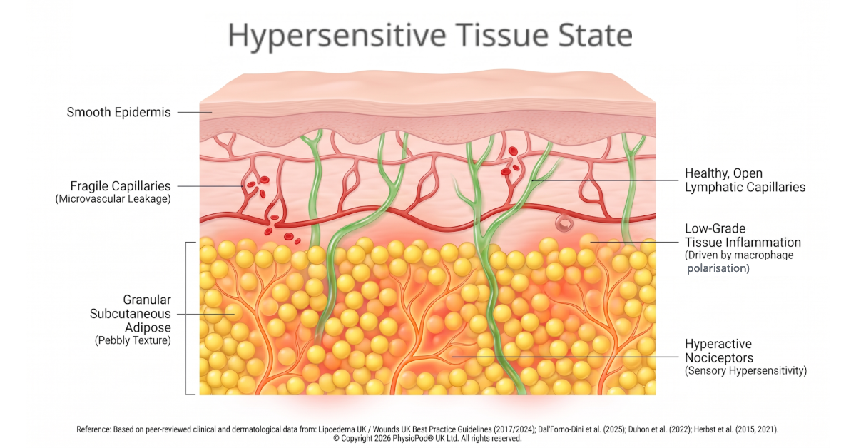
Figure 4: The hypersensitive tissue state - A microscopic cross-section outlining the biology of tissue hypersensitivity © Copyright 2026 PhysioPod® UK Ltd. All rights reserved.
- The Biology: Subcutaneous low-grade inflammation, connective tissue matrix fragility, and hyperactive pain receptors (nociceptors) drive easy bruising, heavy limbs, and intense pressure sensitivity (Dal'Forno-Dini et al., 2025). Skin appears smooth but has a soft, granular or "pebbly" texture beneath on palpation (Herbst et al., 2015).
- In-Clinic Care (MLD & Deep Oscillation): The clinician combines Manual Lymphatic Drainage (MLD) with Deep Oscillation, utilising light vinyl gloves as the therapy's medium. Operating on a pulsed electrostatic field that generates gentle endogenous tissue microdeformations through repeated cycles of tissue attraction and release (Hernández Tápanes et al., 2018), this approach comfortably soothes overstimulated pain pathways (Boisnic & Branchet, 2013). This allows the therapist to deliver highly effective pain-relieving care without applying painful downward physical pressure on fragile, tender tissues.
- Self-Care at Home: For regular conservative maintenance between clinic visits, patients self-treat using a portable Deep Oscillation device. Gliding a handheld applicator (or applicators) over the skin contributes to pain modulation and improved tissue comfort with minimal physical pressure (Hernández Tápanes et al., 2018), helping to comfortably manage daily tenderness, reduce localised tissue stress, and restore physical comfort with minimal effort.
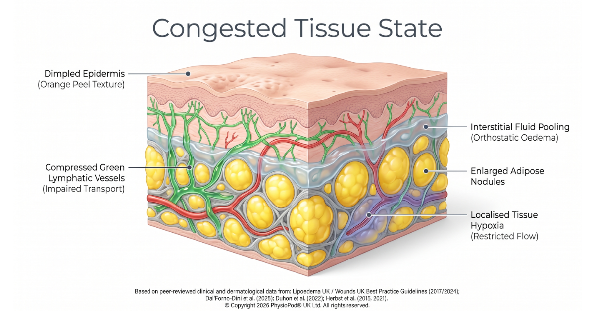
Figure 5: The congested tissue state - A microscopic cross-section outlining the biology of congested tissue
© Copyright 2026 PhysioPod® UK Ltd. All rights reserved.
- The Biology: Where localised tissue stress or fluid clearance fluctuates, metabolic and lymphatic waste products can pool in the interstitial spaces, leading to localised tissue swelling (orthostatic oedema), skin temperature shifts (affected limbs feeling noticeably cooler), and a deep-seated sensation of aching tightness (Dal'Forno-Dini et al., 2025). Symmetrical subcutaneous fat pads around the inner knees can also become larger, heavier, and increasingly sensitive.
- In-Clinic Care (MLD & Deep Oscillation): Rhythmic attraction and release settings are introduced to comfortably permeate deeper tissue planes. This serves as a supportive clinical adjunct to standard MLD, assisting the therapist in comfortably reabsorbing stagnant, stubborn fluid and softening localised nodular congestion (Teo et al., 2016) using incredibly light, non-traumatic movements.
- Self-Care at Home: Daily self-treatment using portable Deep Oscillation devices (Nourollahi et al., 2013) helps patients proactively manage orthostatic swelling, clearing fluid build-up in the evenings and preventing daily limb tightness and heaviness from settling.
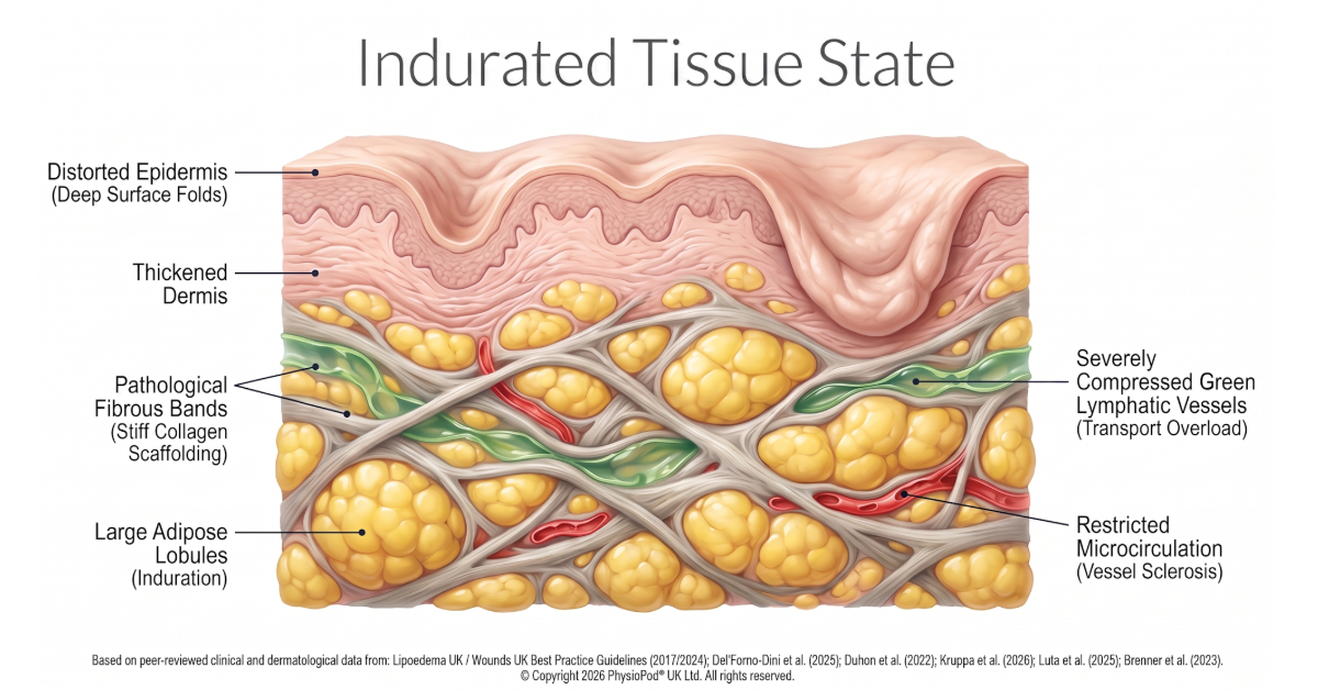
Figure 6: The indurated tissue state - A microscopic cross-section outlining the biology of indurated tissue
© Copyright 2026 PhysioPod® UK Ltd. All rights reserved.
- The Biology: In tissues characterised by dense, established structural changes, or where localised tension has accumulated over time, the chronic low-grade inflammatory state can trigger subcutaneous tissue thickening (pathological fibrosis) (Faerber et al., 2024). Connective tissue fibres can become dense, stiff, and lose their natural elasticity, forming hard, palpable nodules that can limit mobility, restrict joint movement, and alter walking gait (Dal'Forno-Dini et al., 2025).
- In-Clinic Care (MLD & Deep Oscillation): Clinicians target these tough, deep-seated fibro-fatty networks using deep-acting, low-frequency pulses. This comfortable, low-frequency mechanical pacing acts to support tissue softening, improve tissue pliability, and reduce stiffness (Hernández Tápanes et al., 2018) in rigid subcutaneous zones that standard manual therapies cannot easily reach.
- Self-Care at Home: Integrating low-frequency portable settings at home allows patients to actively maintain these clinical gains. Regular, self-directed use helps prevent tissue hardening and maintains joint comfort, which is essential for preserving joint range of motion, protecting walking posture, and maintaining daily mobility (Hernández Tápanes et al., 2018)
8. How Deep Oscillation interacts in the tissue layers
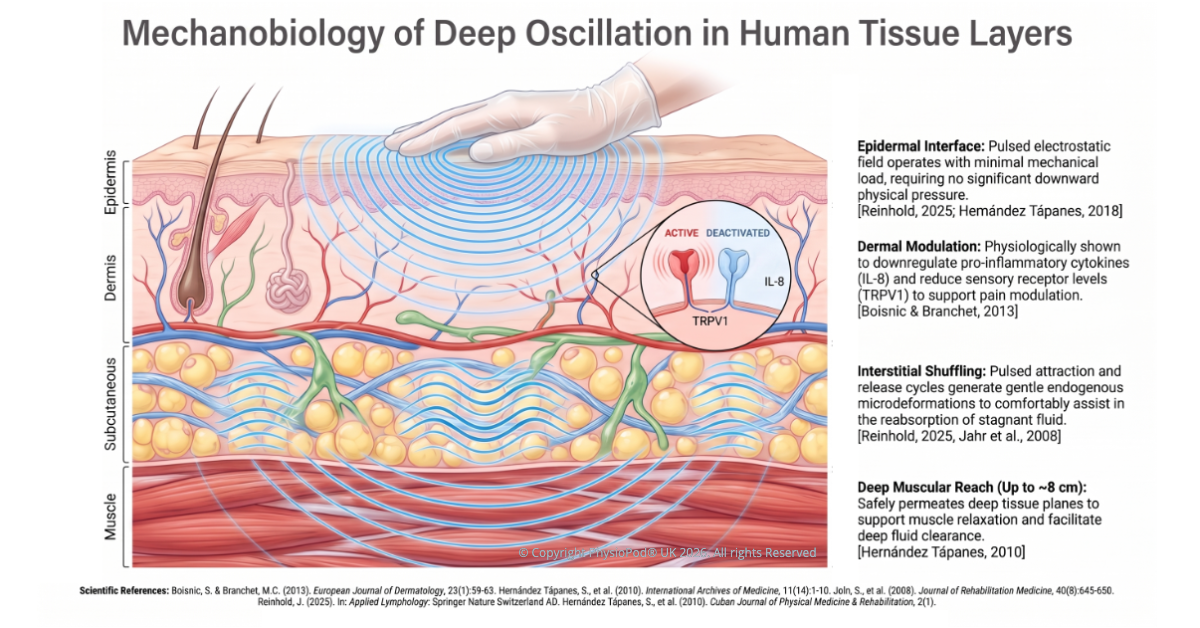
Figure 7: Mechanobiology of Deep Oscillation in Human Tissue Layers - A 3D vertical anatomical schematic of the skin, subcutaneous fat, and muscle layers under the influence of a pulsed electrostatic field (the Johnsen-Rahbek effect). The diagram illustrates pressure-free epidermal contact, dermal cytokine and sensory receptor downregulation, interstitial shuffling of the extracellular matrix, and deep muscular permeation up to 8 cm. On-image annotations highlight peer-reviewed clinical data and laboratory verifications. © Copyright 2026 PhysioPod® UK Ltd. All rights reserved.
- Epidermal & Dermal Skin Layers: Soothing overstimulated pain receptors and downregulating local inflammatory responses (Boisnic & Branchet, 2013).
- Interstitial Fascial Networks: Keeping interstitial septa and tissue pathways open to support the natural reabsorption of stagnant fluid (Almendras et al., 2024).
- Subcutaneous Adipose Tissue: Softening consolidated tissue structures and improving tissue pliability (Hernández Tápanes et al., 2018).
- Deep Muscle & Lymphatic Structures: Promoting muscle relaxation, tissue trophicity, and microcirculation (Boisnic & Branchet, 2013).
Mechanobiological Evidence: Laboratory & Ex-Vivo Proof
- Dermal Inflammation & Capillary Dilation: The study demonstrated that the therapy supported the reduction of local tissue inflammation, showing a reduction in dilated capillaries and a downregulation of pro-inflammatory cytokines, specifically Interleukin-8 (IL-8) (Boisnic & Branchet, 2013).
- Sensory Receptor Modulation: Histological and immunohistochemical analysis indicated that the biophysical interaction significantly lowered sensory receptor levels (specifically TRPV1) in the dermal layers (Boisnic & Branchet, 2013). This research helps explain how the therapy supports pain modulation and contributes to improved tissue comfort.
- Interstitial Shuffling of the Basic Substance: On the level of the interstitium, the rhythmic attraction and release cycles are described as inducing a physical "shuffling" of the basic substance (Reinhold, 2025). This biophysical interaction helps maintain the patency of delicate interstitial septa and fissures, supporting natural fluid clearance, assisting in the reabsorption of stagnant oedema (Almendras et al., 2024), and helping to limit the development of dense, hard fibrotic tissue.
Video 1: Deep Oscillation Theory & Visual Evidence - Begins with a short video of Dr Marianne Lou, a specialist in Oncological and Dermatofunctional Physiotherapy, demonstrating the gentle application of intermittent electrostatic impulses via gloved hands in Lipoedema to generate gentle endogenous microdeformations, comfortably supporting pain modulation, tissue pliability, and lymphatic drainage. © Copyright 2026 PhysioPod® UK Ltd.
9. Supporting connective tissue: direct and extrapolative clinical evidence
10. Surgical integration: supporting pre- and post-operative pathways
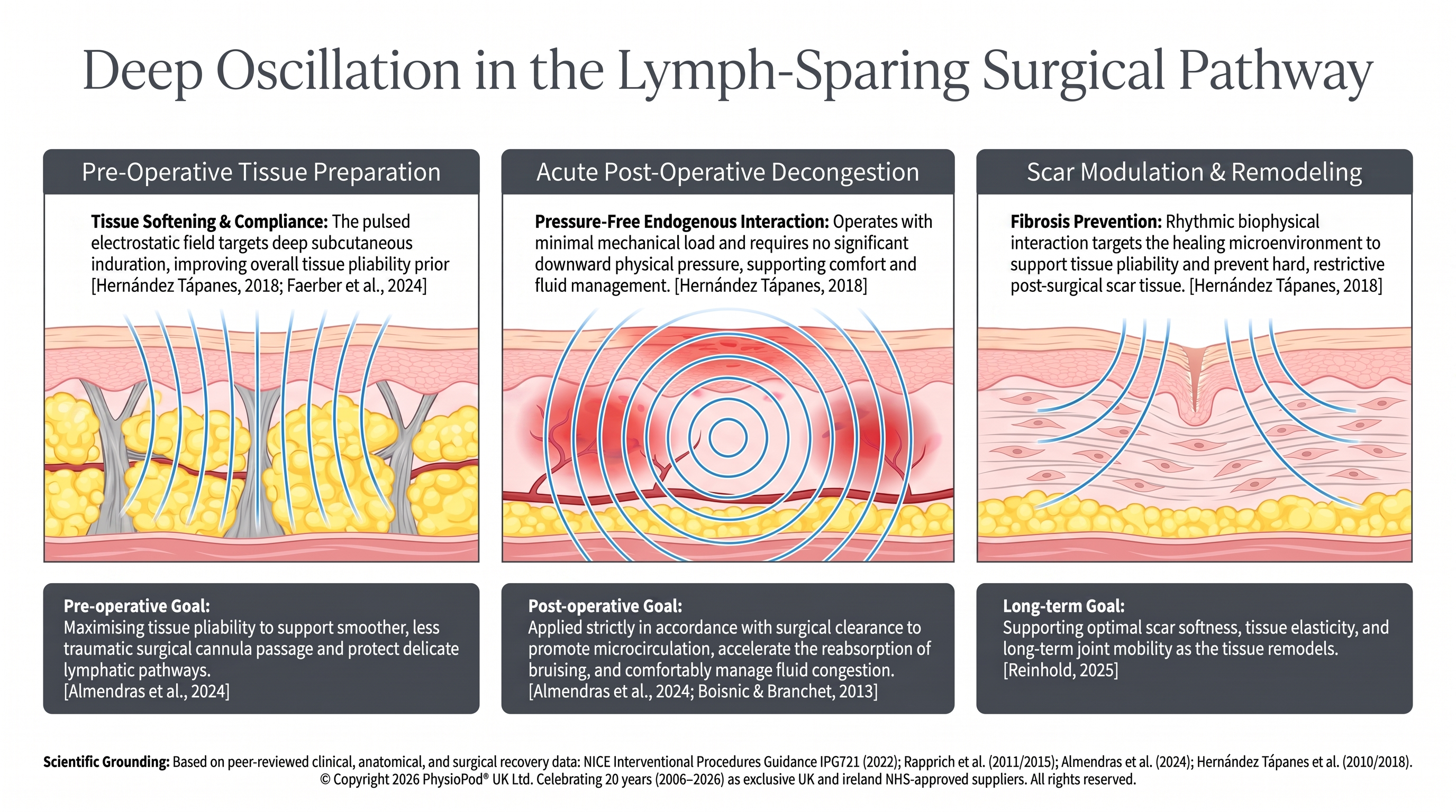
Figure 8: Deep Oscillation in the Surgical Pathway - A clinical schematic illustrating pulsed electrostatic support across the pre-operative, acute post-operative, and tissue-remodelling phases. The diagram demonstrates pre-operative support for tissue softening, pressure-free post-operative fluid clearance, and long-term scar remodelling to preserve tissue pliability and joint mobility—all operating with minimal mechanical load and requiring no downward physical contact pressure. © Copyright 2026 PhysioPod® UK Ltd All rights reserved
11. Combining therapies for daily life
- Hydrostatic Compression: The natural pressure of water exerts a gentle, uniform hydrostatic pressure gradient on the limbs. This supports venous and lymphatic return and assists in the clearance of stagnant fluid without applying painful physical pressure (Wounds UK, 2017).
- Buoyancy and Joint Relief: Water supports up to 90% of body weight, significantly reducing gravitational strain on hypermobile hips, knees, and ankles. This supports a wider, more comfortable range of joint motion to help support the surrounding muscle pump.
- Tissue Pliability: Regular aquatic movement, including specialised activities like aqua-cycling, has been shown to support tissue pliability and assist in softening subcutaneous tissue in the legs and arms (HSE, 2026).
- Conservative Physical Care: Complex Decongestive Therapy (CDT)—combining professional manual lymphatic drainage (MLD/SLD), medical-grade custom compression garments, daily skincare, and water-based physical activities (HSE, 2026).
- Targeted Metabolic & Surgical Support: Integrating highly individualised nutritional strategies (such as anti-inflammatory or lower-carbohydrate eating) alongside specialist, lymph-sparing surgical interventions when conservative self-management pathways require further reinforcement (Faerber et al., 2024).
- Essential Emotional Support: Accessing specialised professional counselling, peer support networks, or cognitive behavioural therapy (CBT) to help process appearance-related distress, navigate medical scepticism, and alleviate the mental and social burden of living with a chronic disease (Wounds UK, 2017).
12. Clinical glossary of biophysical and medical terms
Adipocytokine Signature
- Everyday Translation: Fat-cell chemical markers
- Scientific Definition: A specific pattern of chemical signals in your blood (specifically FGF21 and chemerin) that could help identify lipoedema, representing a major development toward a reliable, objective diagnostic test.
- Clinical Evidence: Mühlberg et al., 2026
Adipose Heterogeneity
- Everyday Translation: Different types of fat tissue
- Scientific Definition: The understanding that fat behaves differently across various parts of your body (such as your arms, legs, or tummy). Each area has its own genetic programme and works like a distinct mini-organ. Note: While a subject of significant academic interest, this investigational concept is not yet clinically conclusive.
- Clinical Evidence: Reid et al., 2026
Complex Decongestive Therapy (CDT)
- Everyday Translation: Combined physical therapy
- Scientific Definition: A standardised, non-surgical treatment programme to support lymphatic and tissue health. It combines professional manual lymphatic drainage (MLD/SLD), custom medical-grade compression, daily skincare, and gentle exercise.
- Clinical Evidence: Wounds UK, 2017
Congested Tissue State
- Everyday Translation: Stagnant fluid buildup
- Scientific Definition: A pathological state where localised tissue fluid clearance fluctuates, causing metabolic and lymphatic waste products to pool in interstitial spaces. This triggers symmetrical swelling (orthostatic oedema), skin temperature cooling, and an aching tightness (often concentrated around knee fat pads).
- Clinical Evidence: Dal'Forno-Dini et al., 2025 | Teo et al., 2016 | Luta et al., 2025
Connective Tissue Matrix Fragility
- Everyday Translation: Weak supportive tissue
- Scientific Definition: An underlying structural weakness in the mesh-like tissue that supports your skin and fat cells. Along with mild inflammation, this fragility can cause easy bruising, heavy limbs, and tenderness.
- Clinical Evidence: Duhon et al., 2022
Cuffing (or Bracelet) Effect
- Everyday Translation: Tissue cuffing at wrists and ankles
- Scientific Definition: A key visual marker of lipoedema where the abnormal fat stops abruptly just above your wrists or ankles, completely sparing your hands and feet.
- Clinical Evidence: Lipedema Foundation, 2025
Endogenous Microdeformations
- Everyday Translation: Inner tissue vibrations
- Scientific Definition: Tiny, microscopic movements and changes in tension generated within your tissues by a pulsed electrostatic field, helping to soothe tissues from the inside out without applying heavy pressure.
- Clinical Evidence: Hernández Tápanes et al., 2018
Extracellular Matrix (ECM) Remodelling
- Everyday Translation: Structural tissue rebuilding
- Scientific Definition: The biological process of rebuilding the supportive scaffolding around your cells. In lipoedema, this scaffolding remodels in a way that can lead to tissue congestion and tightness.
- Clinical Evidence: Duhon et al., 2022
Glycaemic Peaks
- Everyday Translation: Blood sugar spikes
- Scientific Definition: Quick, sharp rises in your blood sugar levels after eating refined carbohydrates. These can trigger insulin spikes and contribute to tissue swelling and inflammation.
- Clinical Evidence: Di Renzo et al., 2021
Gynoid Adipose (Female-Pattern Fat)
- Everyday Translation: Lower-body fat
- Scientific Definition: Subcutaneous fat that naturally settles on the hips, buttocks, and thighs. Unlike abdominal fat, lower-body fat acts as a "metabolic shield" that supports heart health. Lipoedema specifically targets these areas.
- Clinical Evidence: Torre et al., 2018
Hypersensitive Tissue State
- Everyday Translation: Tender, easily bruised skin
- Scientific Definition: A tissue presentation characterised by subcutaneous low-grade inflammation, extracellular matrix fragility, and hyperactive nociceptors (pain receptors). This results in intense pressure sensitivity, mechanical tenderness, and a granular ("pebbly") subcutaneous texture on palpation.
- Clinical Evidence: Dal'Forno-Dini et al., 2025 | Herbst et al., 2015 | Boisnic & Branchet, 2013
Indurated Tissue State
- Everyday Translation: Firm, stiffened tissue
- Scientific Definition: An advanced tissue state where chronic low-grade inflammation triggers dense subcutaneous tissue thickening and pathological fibrosis. Connective tissue fibres lose their natural elasticity and form hard, palpable nodules that restrict joint range of motion and joint compliance.
- Clinical Evidence: Faerber et al., 2024 | Dal'Forno-Dini et al., 2025 | Hernández Tápanes et al., 2018
Johnsen-Rahbek Effect
- Everyday Translation: Electrostatic attraction and relea
- Scientific Definition: The biological science behind Deep Oscillation® therapy. A gentle electrostatic field creates a rhythmic "attraction and release" effect between the therapist's vinyl-gloved hands (or applicator) and your skin.
- Clinical Evidence: Hernández Tápanes et al., 2018
Lipo-lymphoedema
- Everyday Translation: Secondary fluid buildup
- Scientific Definition: A secondary buildup of fluid that happens when otherwise healthy lymphatic pathways are squeezed or stressed by heavy lipoedema tissue or extra weight. It is not an inevitable stage of the condition.
- Clinical Evidence: Shavit & Wollina, 2018 | HSE Consensus Guideline, 2026
Macrophage Polarisation
- Everyday Translation: Overactive immune cells
- Scientific Definition: A process where your body's protective immune cells switch into an overactive, inflammatory state. This can drive chronic, low-grade tissue inflammation and changes in fat tissue.
- Clinical Evidence: Duhon et al., 2022
Nociceptors
- Everyday Translation: Pain-sensing nerves
- Scientific Definition: Specialised nerve endings in your tissues that send pain signals to your brain. Low-grade tissue inflammation can make them highly sensitive, but managing them helps soothe discomfort.
- Clinical Evidence: Dal'Forno-Dini et al., 2025
Psychonutrition
- Everyday Translation: Mind-food connection
- Scientific Definition: A clinical field that looks at how your food choices affect your emotions and brain health. It helps support patients in managing food-related anxiety and building a balanced, nourishing relationship with food.
- Clinical Evidence: Kruppa et al., 2026
Reddit r/lipedema Community
- Everyday Translation: Online peer support
- Scientific Definition: A global online forum where women share their real-life experiences of lipoedema, helping to validate symptoms like widespread tissue ache that lie outside rigid clinical checklists.
- Clinical Evidence: Reddit r/lipedema
Stemmer's Sign
- Everyday Translation: Skin-pinch test
- Scientific Definition: A simple physical test where a practitioner tries to pinch a fold of skin at the base of your second toe. If they can pinch it (negative sign), it points to lipoedema rather than lymphoedema.
- Clinical Evidence: Shavit & Wollina, 2018
Tissue Induration
- Everyday Translation: Firm, hardened tissue
- Scientific Definition: The physical thickening and stiffening of your under-skin tissues, which can occur when fluid congestion and fibrous networks build up over a long period.
- Clinical Evidence: Hernández Tápanes et al., 2018
Tissue Trophicity
- Everyday Translation: Tissue nourishment
- Scientific Definition: The overall state of nutrition and cell health in your tissues. This is closely linked to healthy blood circulation and oxygen flow, which help protect and support your tissue layers.
- Clinical Evidence: Boisnic & Branchet, 2013
Waist-to-Height Ratio (WHtR)
- Everyday Translation: Waist-to-height ratio
- Scientific Definition: A simple health measurement calculated by dividing your waist size by your height. It is highly recommended over BMI in lipoedema because it measures central health while ignoring limb weight.
- Clinical Evidence: Brenner et al., 2023
Waist-to-Hip Ratio (WHR)
- Everyday Translation: Waist-to-hip ratio
- Scientific Definition: A comparison of your waist and hip sizes. In women, a lower score (under 0.70 to 0.75) shows that tissue is concentrated in the lower limbs, helping clinicians assess lipoedema.
- Clinical Evidence: HSE Consensus Guideline, 2026
13. References
Note: This bibliography serves as a comprehensive reference library for both the inline citations in this article and the primary literature featured in the downloadable companion resource, the Lipoedema Scientific Journey Timeline (1940–2026) PDF
Allen, E. V., & Hines, E. A. (1940). “Lipoedema of the legs: a syndrome characterised by fat legs and orthostatic edema.” Proceedings of the Staff Meetings of the Mayo Clinic, 15, pp. 184–187. (Note: This historic foundational paper is not digitised).
Almendras, M. J., et al. (2024). “The effects of shock waves and deep oscillations on lipoedema.” Journal of Aesthetic Nursing, 13(2), pp. 42–53. [Paper Link]
Boisnic, S., & Branchet, M. C. (2013). “Anti-inflammatory and draining effect of the Deep Oscillation® device tested clinically and on a model of human skin maintained in survival condition.” European Journal of Dermatology, 23(1), pp. 59–63. [Paper Link]
Brenner, E., et al. (2023). “Body mass index vs. waist-to-height-ratio in patients with lipohyperplasia dolorosa (vulgo lipedema).” Journal der Deutschen Dermatologischen Gesellschaft (JDDG), 21(10), pp. 1179–1185. [Paper Link]
Cannataro, R., et al. (2021). “Management of Lipedema with Ketogenic Diet: 22-Month Follow-Up.” Life, 11(12), p. 1402. [Paper Link]
Chachaj, A., et al. (2023). “Lymphoscintigraphic alterations in lower limbs in women with lipedema in comparison to women with overweight/obesity.” Frontiers in Physiology, 14, p. 1099555. [Paper Link]
Child, A., et al. (2010). “First Genetic Inheritance Mapping of Lipoedema.” American Journal of Medical Genetics. [Paper Link]
Cifarelli, V. (2025). “Lipedema: Progress, Challenges, and the Road Ahead.” Obesity Reviews, 26(10), p. e13953. [Paper Link]
Daia, C. O., Paduraru, D. N., Radu, L., Lipianu, I., & Bumbea, A. M. (2026). “Electrotherapy in Oncology Rehabilitation: Current Evidence, Safety Considerations, and Future Perspectives.” Journal of Clinical Medicine, 15(14), 5548. [Paper Link]
Dal'Forno-Dini, T., et al. (2025). “Lipedema: pathophysiological insights and therapeutic strategies – An update for dermatologists.” Anais Brasileiros de Dermatologia. [Paper Link]
Di Renzo, L., et al. (2021). “Potential Effects of a Modified Mediterranean Diet on Body Composition in Lipoedema.” Nutrients, 13(2), p. 358. [Paper Link]
Duhon, B. H., et al. (2022). “Current Mechanistic Understandings of Lymphedema and Lipedema: Tales of Fluid, Fat, and Fibrosis.” International Journal of Molecular Sciences, 23(12), p. 6621. [Paper Link]
Faerber, G., et al. (2024). “S2k guideline lipedema.” Journal der Deutschen Dermatologischen Gesellschaft (JDDG), 22(9), pp. 1303–1315. [Paper Link]
Fedre, B., Dessalvi, S., and Boccardo, F. (2025). “A Case Series on Combining Modified Mediterranean Diet and Ketogenic Diet in a “Sandwich” Approach for Patients with Lipedema and Comorbidities.” Lymphology, 58(3), pp. 108–118. [Paper Link].
Health Service Executive (HSE). (2026). “HSE Consensus Guideline for the Diagnosis and Non-Surgical Management of Lipoedema/Lipalgia Syndrome (LLS).” National Lymphoedema Oversight Team. [Paper Link]
Hyden, L. C. (1997). “Illness and narrative.” Sociology of Health & Illness, 19(1), pp. 48–69. [Paper Link]
Herbst, K. L., et al. (2015). “Lipedema Fat and Signs and Symptoms of Illness, Increase with Advancing Stage.” Archives of Medicine, 7(4), p. 10. [Paper Link]
Herbst, K. L., et al. (2021). “Standard of care for lipedema in the United States.” Phlebology: Journal of Venous Disease, 36(10), pp. 779–796. [Paper Link]
Hernández Tápanes, S., Socas Fernández, M. D. J., Iturralde, Y., & Suáres Fernández, A. (2018). “The Effect of Deep Oscillation Therapy in Fibrocystic Breast Disease: A Randomised Controlled Clinical Trial.” International Archives of Medicine, 11(14), pp. 1–10. [Paper Link]
Kruppa, P., et al. (2026). “Lipedema World Alliance Delphi Consensus-Based Position Paper on the Definition and Management of Lipedema: Results from the 2023 Lipedema World Congress in Potsdam.” Nature Communications, 17(1), p. 427. [Paper Link]
LaMantia, J. (2024). “A Clinical Guide to Managing GLP-1 Side Effects and Preserving Muscle in Lipoedema.” [Paper Link]
Lipedema Foundation. (2025). “About Lipedema: Diagnostic Features and Sparing.” [Web Link]
Lipoedema UK. (2026). “Lipoedema Stages & Types – Lipoedema Classification Explained.” [Web Link]
Luta, X., et al. (2025). “Clinical characteristics, comorbidities, and correlation with advanced lipedema stages: A retrospective study from a Swiss referral centre.” PLoS One, 20(3), p. e0319099. [Paper Link]
Lundanes, J., et al. (2024). “Effect of a low-carbohydrate diet on pain and quality of life in female patients with lipedema: a randomised controlled trial.” Obesity, 32(6), pp. 1071–1082. [Paper Link]
Medscape. (2026). “Lipedema: A Multidisciplinary and Surgical Approach to Care.” Medscape Medical News, August 6, 2026. Reporting from the International Congress on Obesity (ICO 2026), Mexico City. [Paper Link]
Mühlberg, K. S., et al. (2026). “An adipocytokine signature improves diagnostic accuracy for people living with lipoedema.” British Journal of Dermatology, ljag103. [Paper Link]
National Institute for Health and Care Excellence (NICE). (2022). “Liposuction for chronic lipoedema.” Interventional procedures guidance [IPG721]. [Paper Link]
Nourollahi, S., et al. (2013). “Incorporating Deep Oscillation Therapy into Standard Conservative Care for Lipoedema.” Alternative & Integrative Medicine, 2, p. 1000122. [Paper Link]
Pinelli, R., et al. (2025). “A Case Series on the Efficacy of the Pharmacological Treatment of Lipedema: The Italian Experience with Exenatide.” Clinical Practice, 15(7), p. 128. [Paper Link]
Reid, I., et al. (2026). “Lipedema as a disorder of adipose tissue heterogeneity: insights from single-cell and spatial transcriptomics.” Frontiers in Cell and Developmental Biology, 14, p. 1809914. [Paper Link]
Reinhold, J. (2025). “Deep Oscillation.” In: Cornely M. E. et al. (eds.), Applied Lymphology. Springer Nature Switzerland AG, pp. 340–355. [Paper Link]
Shavit, E., and Wollina, U. (2018). “Lipoedema is not lymphoedema: A review of current literature.” International Wound Journal, 15(6), pp. 921–928. [Paper Link]
StatPearls. (2025). “Lipedema.” StatPearls [Internet]. Treasure Island (FL): StatPearls Publishing. [Paper Link]
Straub, L. G., et al. (2025). “Defining lipedema's molecular hallmarks by multi-omics approach for disease prediction in women.” Metabolism, 168, p. 156191. [Paper Link]
Teo, T., Coulborn, A., & Munnoch, D. A. (2016). “Use of the DEEP OSCILLATION® system in the treatment of secondary lymphoedema of the breast.” Journal of Lymphoedema, 11(1), pp. 13–17. Paper Link
Torre, Yanira Sanchez-De la, Wadeea, Rita, Rosas, Victoria and Herbst, Karen L.. "Lipedema: friend and foe" Hormone Molecular Biology and Clinical Investigation, vol. 33, no. 1, 2018, pp. 20170076. [Paper Link]
Tomiyama, A. J., Mann, T., Vinas, D., et al. (2010). “Low calorie dieting increases cortisol.” Psychosomatic Medicine, 72(4), pp. 357–364. [Paper Link]
Verde, L., Camajani, E., Annunziata, G., et al. (2023). “Ketogenic Diet: A Nutritional Therapeutic Tool for Lipedema?” Current Obesity Reports, 12, pp. 529–543. [Paper Link].
Wounds UK. (2017). “Best Practice Guidelines: The Management of Lipoedema.” London: Wounds UK. [Paper Link]
14. Imprint, attribution and transparency statement
- Bel Hardman (Founder, Patient Advocate, Lipedema Circle): For her deeply compassionate, patient-first review of this work, her visual design suggestions, and her tireless advocacy in shifting the focus of the lipoedema community toward deep cellular biology and sustainable, individualised care.
- Jacqueline (Jax) Smith (Patient Advocate): For her somatic, lived-experience insights, her invaluable feedback on joint displacement and gravitational fluid dynamics, and her dedication to keeping the patient's voice at the absolute heart of this clinical review.
- Dr Solangel Hernández Tápanes, MD, PhD (Specialist PM&R Physician & President of AMLAR): For her pioneering research and for developing The Hernández Model of Tissue-Centred Clinical Reasoning that forms the heart of our clinical philosophy.
- Dr Garry Cooper, QN, RGN (National Research & Innovation Lymphoedema Specialist): For sharing his clinical expertise, meticulously reviewing our physical medicine guidelines, and helping us align our writing with the latest medical consensus.
- Dr Anne Williams, PhD, RGN (Nurse Consultant & Research Advisor): For generously reviewing our draft, guiding our sensitive reframing of the lipoedema staging debate, and helping us clinically anchor our nutritional cortisol and Wounds UK guideline references.
- Christine Talbot (Specialist Lymphoedema Practitioner): For her outstanding attention to detail, expert proofreading, and total support of all our endeavours.
- Catherine Groom (Specialist Lymphoedema Practitioner): For her exceptional clinical insights, meticulous reviews of our therapy pathways, and invaluable frontline dedication to reassuring patients anxious about disease progression.
- Publisher: PhysioPod® UK Ltd, Sherbrook Road, Daybrook, Nottingham, NG5 6AS.
- Tel: +44 (0)115 916 7685 | Email: enquiries@physiopod.co.uk | Web: www.physiopod.co.uk.
